club foot horse x ray
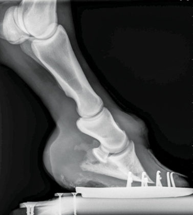
Club foot is defined as a flexural deformity of the coffin joint and is a common problem in young growing horses. Assess the whole horse not just the hooves and identify and treat any potential problems above the hoof first.
Understanding Navicular Syndrome Heel Pain In Horses
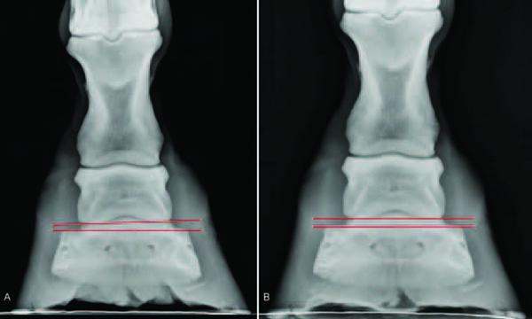
Lateral talocalcaneal angle.
. There are varying degrees of club foot. Club Foot Heritability in Horses. Home Practice Team Contact 201 806-6099.
Physicians Surgeons Radiology Medical Dental X-Ray Labs Physicians Surgeons 732 390-0030. Physicians Surgeons Radiology Physicians Surgeons Medical Dental X-Ray Labs. If the axis is broken forward club foot or if the axis is broken back long toe.
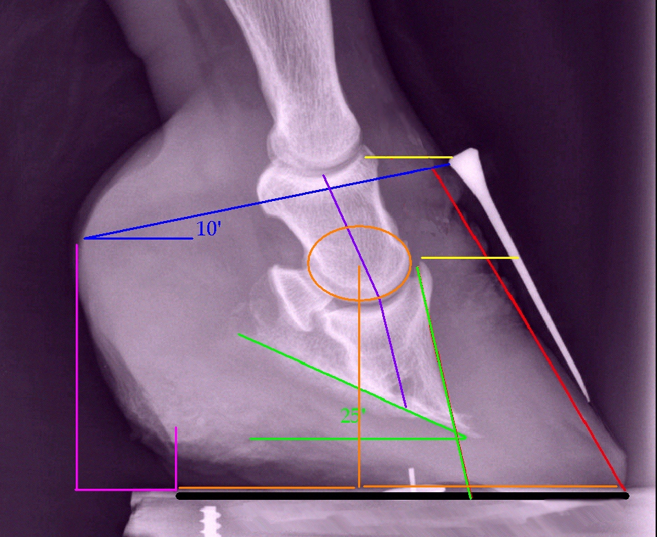
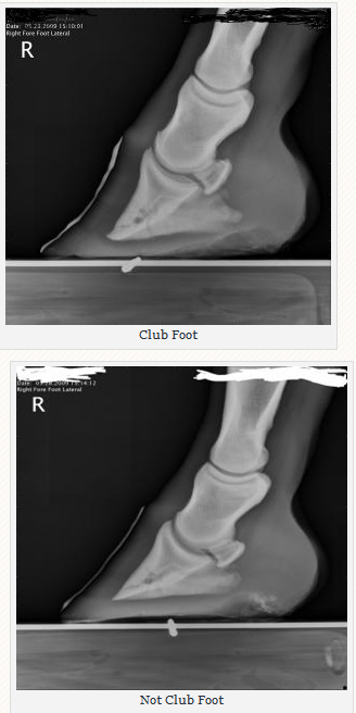
The normal range of hoof angle is 50 to 55 degrees while a club foot might stand at more than 60 degrees. 33 Central Ave Midland Park NJ 07432. An X ray of your horses foot can help you predict the future while it shows you the present.
The professional services are ready to your most urgent injuries form lots of bones to compound fractures in the Radiology Center at Harding X-ray services can help. The x-ray will show whether the hoof pastern axis is parallel. A normal angle for a horses hooves varies by the individual.
The condition is most often encountered in young. When the hoof angle of one foot is 3 to 5. On sound horses hooves tend to.
Medial subluxation of the navicular on the talus. Stretching massage therapy and chiropractic treatments all help increase the range of motion and along with trimming can play an important role in the maintenance prevention and even the correction of club feet. Website Services 732 632-1650.
In a club foot the angle of the hoof and pastern in relation to the ground is abnormally steep. Many people call the ahead using services as well as wait in the lobby for your X-Ray. Talipes equinovarus consists of four elements 7.
Lateral radiograph of the right foot shows that the long axes of the talus and calcaneus are nearly parallel. Peterson Mary A MD. Equine club foot has several distinguishing characteristics Don says.
A matter of degree. It is more available from the need to X-ray for any type of injury with the get checked out. East Brunswick NJ 08816.
MedWell Testosterone Weight Loss Hormone Replacement 201 806-6099. 33 Central Ave Midland Park NJ 07432 201 806-6099. Apply to X-ray Technician Technician Sterilization Technician and more.
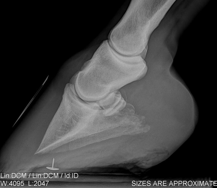
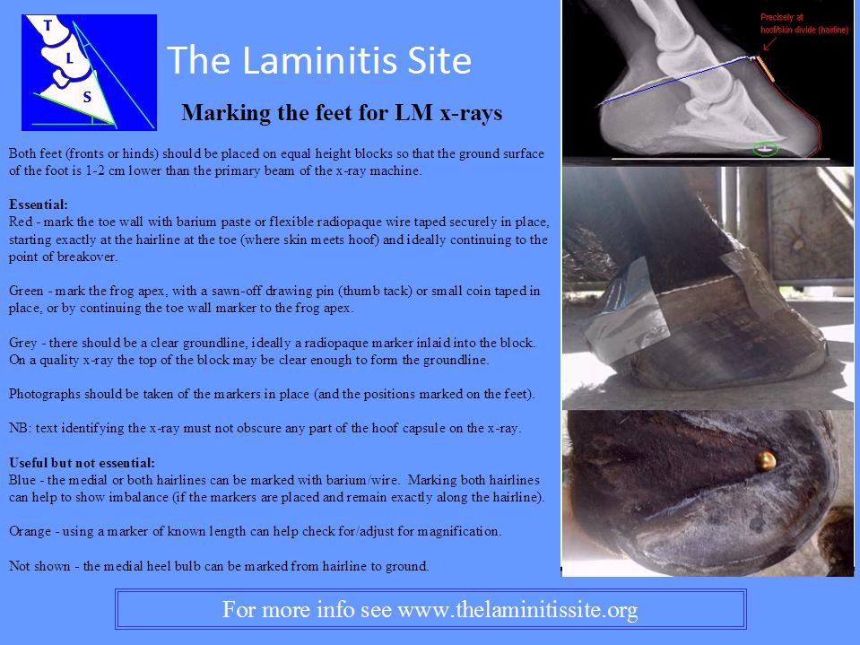
Radiographic evaluation of the dorsal wall with a conforming marker allows accurate assessment of the. As the foot grows out in these horses there is a propensity for the dorsal wall to distort and flare producing multiple angles to the dorsal wall. MECHANICAL LAMINITIS TREATMENT.
The normal alignment of the short pastern bone and coffin bone is a straight line visible on X ray but. In the past the condition was defined as any hoof angle that exceeded 60 degrees but the reality is not quite that exact. Talus to first metatarsal Mearys angle 15º.
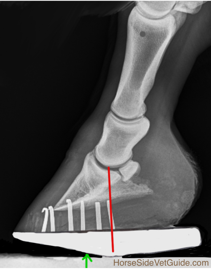
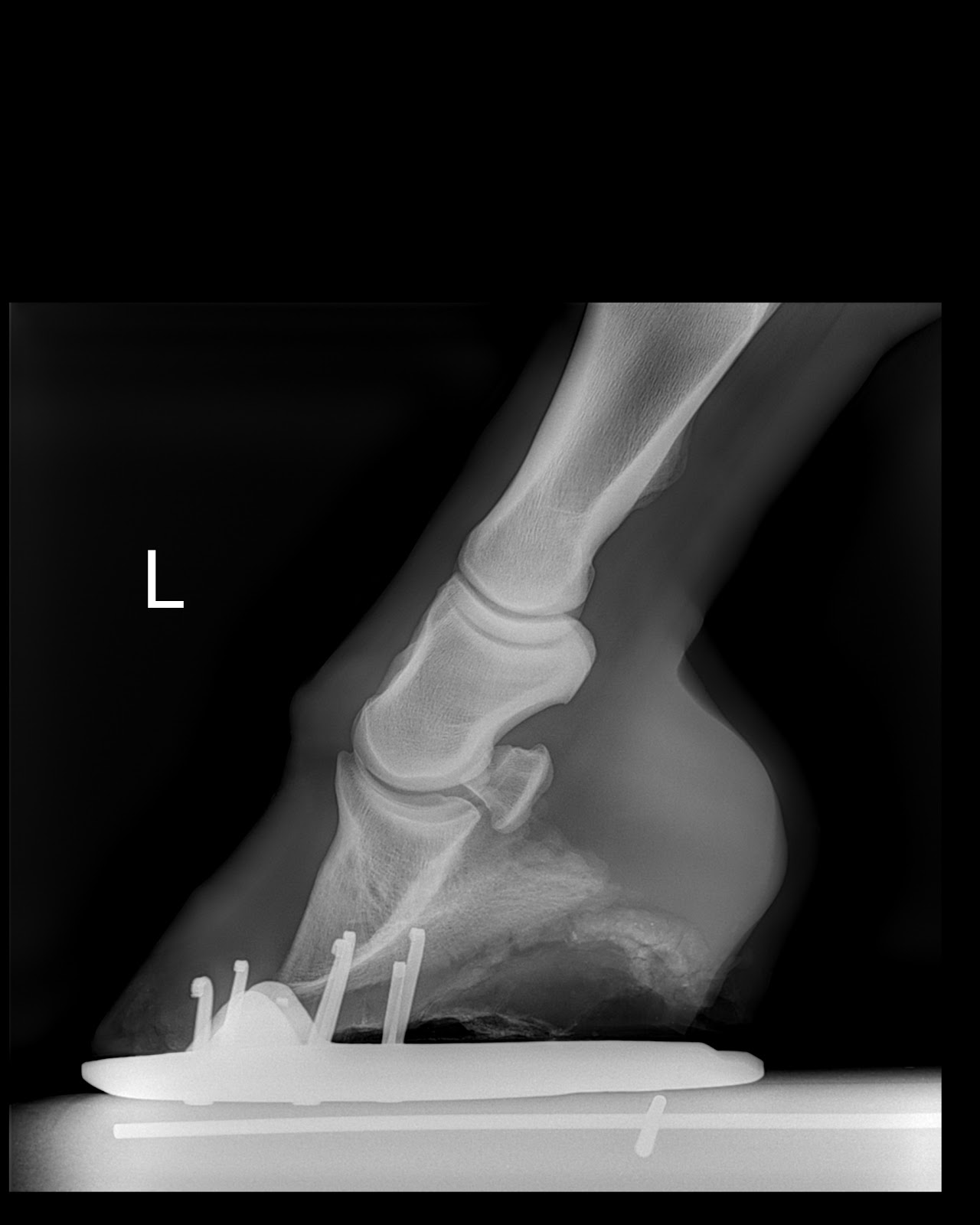
To appreciate bone position the radiographs should be taken with the horse bearing weight and both feet placed on wooden blocks of equal height. In the club foot because the deep flexor tendon is contracted the xray will show that the pedal bone angles are quite different the front is not in line with the hoof wall the tip is pointing down and the rear part is much greater than five degrees Put simply the heels will need to be lowered and any flare corrected at the toeAlmost always I am called to club foot cases. Generally the greater the upright angle the more severe the club foot.
Comes very useful in horses with upright feet the best example being the club-footed horse. The longitudinal arch is abnormally high. Horses feet are placed on x ray transparent lifts or blocks to enable x rays to show complete hoof including sole.
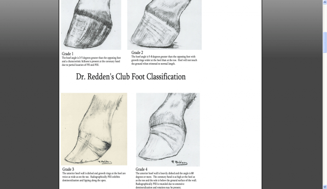
For example lets say you have a grade 2 club foot one thats at least five degrees steeper than the other front or hind foot. Composition main components parts model no. X-Ray providers in Hackensack NJ.
You might see a HL zone of 1516 mm 20 mm of sole and a fairly. The horse should be stood on a flat level surface. AP radiograph of the right foot shows abnormally narrow talocalcaneal angle.
The equine club foot is defined as a hoof angle greater than 60 degrees. What we see externally as the equine clubbed foot is actually caused by a flexural deformity of the distal interphalangeal joint coffin joint. Adduction and varus deformity of the forefoot.
Causes include nutritional issues heredity position in the uterus or injury.
Understanding X Rays The Laminitis Site
2

Hoof Evaluation Radiographs For The Farrier
High Heels The Laminitis Site

Hoof Evaluation Radiographs For The Farrier
Equine Therapeutic Farriery Dr Stephen O Grady Veterinarians Farriers Books Articles

Hoof Evaluation Radiographs For The Farrier

Hoof Radiographs Springhill Equine Veterinary Clinic

How To Treat Club Feet And Closely Related Deep Flexor Contraction
2
Low Foot Case Study Dixie S Farrier Service

The Wild Mustang Hoof

Laminitis A Pictorial Review
Managing The Club Hoof Easycare Hoof Boot News
Understanding X Rays The Laminitis Site

Understanding X Rays The Laminitis Site

The Wild Mustang Hoof
Managing The Club Hoof Easycare Hoof Boot News
Club Foot Or Upright Foot It S All About The Angles